Thyroid US Reader
An AI system that processes thyroid ultrasound studies and generates ACR TI-RADS compliant structured radiology reports. Addresses the most cognitively demanding, time-inefficient task in daily radiology practice.
Research System - Not FDA Cleared. This system is used solely for internal research, development, and technical validation purposes by the founding physician-engineer. It does not constitute a medical device, clinical decision support system, or diagnostic tool. All AI outputs require review and authorization by a licensed physician before any clinical application. VoxelMD is not intended to replace physician judgment. No patient data is processed or stored through this system.
The Clinical Problem
Thyroid ultrasound reporting is one of the highest-volume, lowest-margin, most cognitively demanding tasks in outpatient radiology - and it remains entirely manual.
600K-1M
Thyroid US Studies / Year (US)
CPT 76536 is one of the most frequently ordered outpatient radiology exams, driven by the 19-68% nodule prevalence in adults undergoing cervical imaging.
5 categories
Per Nodule, Per Patient - Manually
ACR TI-RADS requires structured evaluation of composition, echogenicity, shape, margin, and echogenic foci for every nodule - documented in a structured dictation.
$300M+
US Addressable Market
Low-reimbursement, high time-cost studies that reduce radiologist throughput. A shortage of 42,000 radiologists projected by 2050 makes automation a structural necessity.
How It Works
A systematic pipeline solves the image-to-nodule attribution problem inherent to multi-image ultrasound studies - the core challenge that makes thyroid US reporting so time-consuming.
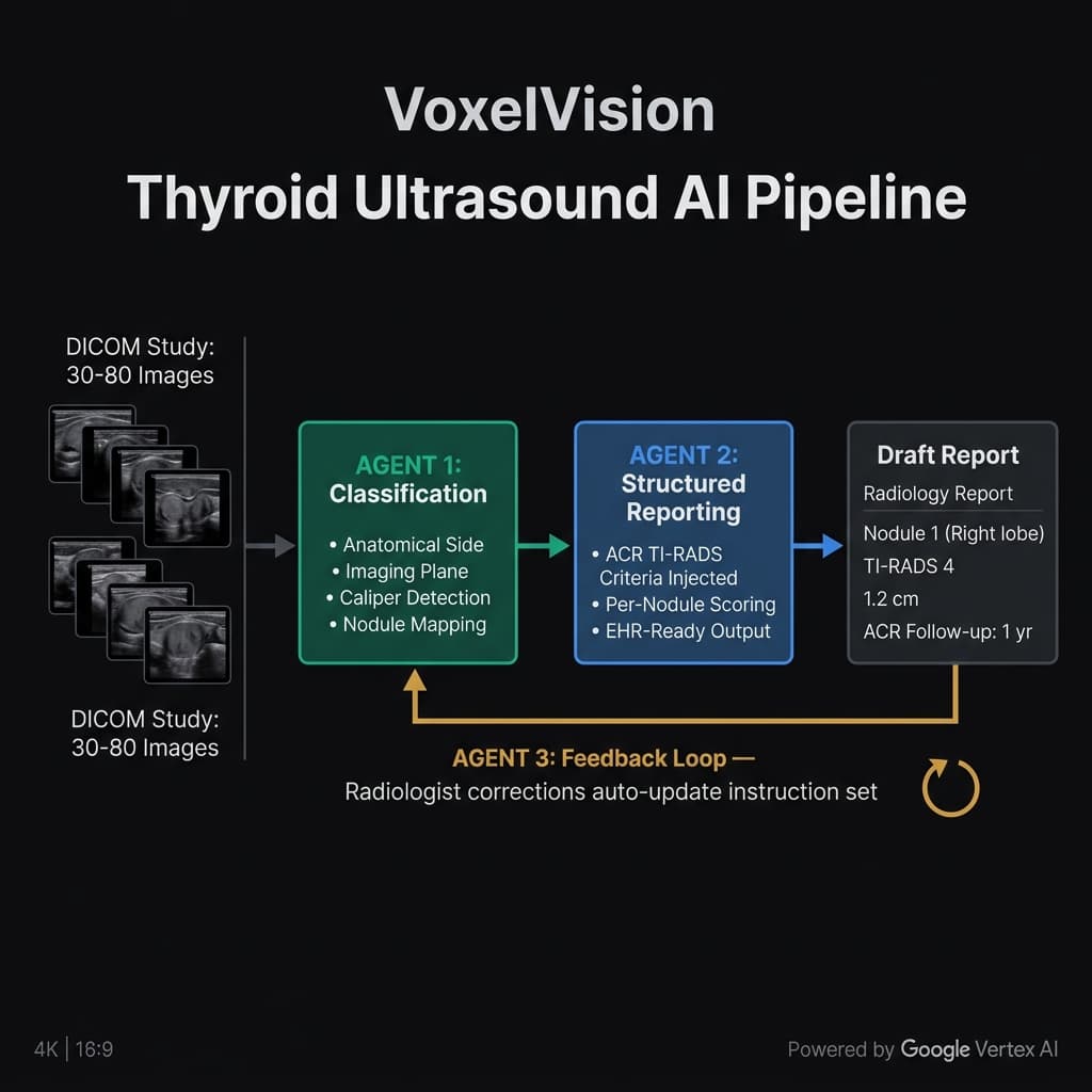
Study Ingestion
A thyroid ultrasound study arrives - typically 30 to 80 DICOM images spanning multiple nodules, bilateral anatomy, and two imaging planes. No metadata maps images to nodules. The only ground truth is the ultrasound machine's text overlay burned into each image's pixel space.
Image Classification - Spatial Mapping
The system parses the burned-in overlay text from each image frame. It identifies anatomical side (right/left lobe), imaging plane (transverse/longitudinal), caliper presence (measurement images), and nodule identity - building a structured nodule-to-image map before any diagnostic reasoning begins. This is the core challenge that makes automated thyroid reporting so difficult.
Structured Reporting - ACR TI-RADS Evaluation
The system ingests the organized, nodule-attributed image sets alongside ACR TI-RADS scoring standards. It evaluates all 5 TI-RADS categories per nodule, requiring explicit image evidence before committing to any feature score - creating an internal audit trail that prevents hallucinated findings.
Structured Report Generation
The system generates a complete field-level structured report: per-nodule TI-RADS scoring, nodule dimensions, laterality, composition, echogenicity, margin classification, echogenic foci, and ACR-compliant follow-up recommendations. Output conforms to EHR data schemas for direct ingestion - not just narrative text.
Continuous Improvement Loop
When a radiologist reviews and corrects the generated report, the system analyzes the correction, identifies the underlying clinical principle, and validates the update against prior cases before incorporating it. The system improves with each correction - building a continuously more accurate engine tuned to each practice.
System in Action
A screen recording demonstrating the VoxelVision thyroid ultrasound reporting pipeline.
Internal research demonstration. No patient data. For technical evaluation purposes only.
Structured Report Output
Screenshots of the VoxelVision interface generating ACR TI-RADS compliant structured reports from live DICOM studies. Each field is AI-populated - composition, echogenicity, margin, echogenic foci, TI-RADS score, and EHR-ready follow-up recommendation.
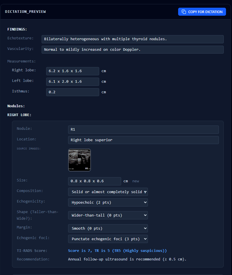
Nodule R1 (Right Lobe Superior) - AI assigned TI-RADS Score 7, TR5 (Highly Suspicious) based on hypoechoic composition, punctate echogenic foci, and caliper-confirmed 0.8 × 0.8 × 0.6 cm dimensions. Source DICOM frame is thumbnailed directly in the report.
EHR-compatible field-level output: bilateral measurements, vascularity, echotexture, per-nodule TI-RADS scoring, and ACR-compliant follow-up recommendation - ready for direct PACS / EHR ingestion.
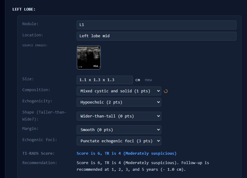
Nodule L1 (Left Lobe Mid) - AI scored TI-RADS 6, TR4 (Moderately Suspicious): mixed cystic and solid composition, hypoechoic, punctate echogenic foci. Follow-up recommendation generated at 1, 2, 3, and 5 years per ACR guidelines.
Multi-nodule studies are handled independently - each nodule receives its own isolated image set, scoring chain, and follow-up recommendation, preventing cross-contamination of findings between anatomical sites.
6 nodules identified across bilateral anatomy, 46 images classified - the system resolved spatial attribution for every image before generating a single field of the report. All output is structured JSON mapped to EHR field schemas, not narrative prose.
Four Core Challenges We Solved
Each challenge is specific to thyroid ultrasound reporting. Each has a defined success metric.
Spatial Mapping Across Multi-Image Studies
Challenge
30-80 images across multiple nodules and bilateral anatomy with no metadata linking images to nodules. A generalist VLM cross-contaminates findings between nodules.
Approach
Dedicated image classification step parses overlay text, identifies caliper presence, and constructs a structured nodule-to-image map before any diagnostic reasoning begins.
Success Metric
≥ 95% image classification accuracy across 500 heterogeneous studies vs. radiologist-annotated ground truth
TI-RADS Hallucination Prevention
Challenge
Evaluating echogenicity or composition in noisy ultrasound images - a VLM is prone to generating plausible but unsupported feature scores without visual evidence.
Approach
The system requires explicit image evidence before committing to any feature score, creating an internal audit trail.
Success Metric
< 2% feature-level hallucination rate across 200 blinded cases, validated by subspecialty radiologist review
EHR-Compliant Structured Output
Challenge
Text that sounds like a radiology report is insufficient. Output must conform to field-level EHR data schemas - TI-RADS score, nodule size, laterality, follow-up encoding - for direct ingestion.
Approach
Constrained output schema enforcement at inference time. Output is field-mapped to EHR schemas, not narrative text. PACS-compatible delivery.
Success Metric
≥ 90% categorical agreement on TI-RADS category and follow-up across 1,000 consecutive de-identified cases; ≥ 85% clinically usable by a panel of 3 subspecialty radiologists
Automated Clinical Rule Extraction
Challenge
The self-improvement loop depends on correctly interpreting what a radiologist correction means clinically. Incorrectly generalizing a correction can degrade performance.
Approach
A dedicated analysis step identifies the clinical principle behind each correction and validates the updated rule against prior cases before incorporation.
Success Metric
Instruction updates that improve concordance on a held-out validation set in ≥ 80% of feedback cases
Self-Improvement in Action
When a physician corrects a report, the system extracts the underlying clinical rule and permanently encodes it - so the system never repeats the same error. These screenshots show the learning loop operating in a live session.
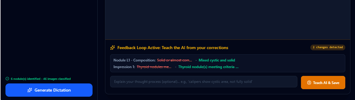
What you're seeing
After reviewing the AI-generated report, two corrections were made: Nodule L1 composition was changed from Solid or almost completely solid → Mixed cystic and solid, and the Impression field was updated. The system detected both changes and surfaces them for physician-guided learning before saving.
The physician can optionally explain their reasoning (e.g. "calipers show cystic area") to improve rule extraction quality before triggering the learning step.
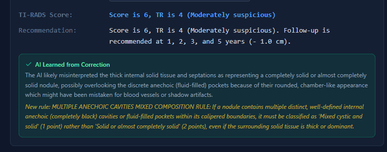
What you're seeing
The system identified why the model erred - it likely misclassified thick internal septations as solid tissue, overlooking the discrete anechoic (fluid-filled) pockets. It then encoded a new permanent clinical rule:
"MULTIPLE ANECHOIC CAVITIES MIXED COMPOSITION RULE: If a nodule contains multiple distinct, well-defined internal anechoic cavities or fluid-filled pockets, classify as 'Mixed cystic and solid' (1 pt) rather than 'Solid or almost completely solid' (2 pts), even if surrounding solid tissue is thick or dominant."
This rule is now applied to every subsequent study - the system will not repeat this classification error for this institution.
Compounding precision: Each correction permanently narrows the model's error surface for that institution's patient population. Unlike static models that plateau after training, VoxelVision's instruction set grows with use - building an institution-specific diagnostic engine that no external competitor can replicate without the same longitudinal correction data.
Cloud Infrastructure
The system runs on Google Cloud for all inference. Medical images are de-identified before any cloud transmission. All proprietary components remain under VoxelMD control.
Phase II Requires
- Advanced model training infrastructure
- Cloud-based fine-tuning capabilities
- High-performance GPU compute for 3D imaging
- Secure storage for training data and models
Market Opportunity
600K-1M
Studies/Year (US)
CPT 76536 thyroid ultrasound
$300M+
Addressable Market
US thyroid US AI reporting
80%
Dictation Time Reduction
Internal estimation
A system that keeps getting better: Each radiologist correction makes the system more accurate for that practice. The longer a group uses VoxelMD, the more accurate and tailored it becomes - a system that keeps getting better.
VoxelMD products are in active research and development. Nothing on this website constitutes a cleared or approved medical device, clinical decision support system, or FDA-regulated software as a medical device (SaMD). No VoxelMD system is cleared or approved by the FDA or any regulatory body for clinical use. All AI outputs are for research and development purposes only and require review and authorization by a licensed physician before any clinical application. Performance metrics referenced are based on internal technical testing and have not been independently validated. Silicon Health Solutions LLC (d/b/a VoxelMD) is a California-registered company.
Explore the Full System
Deployment options, expanding coverage, and product roadmap.