AI Reads the Images.
You Sign the Report.
Voxel Vision drafts complete, structured reports across 25+ exam types in X-ray, ultrasound, and CT, ready the moment the study is opened, inside the radiologist's existing dictation software.
3 relevant priors matched
impressions summarized
Reason for exam captured
shortness of breath
Clinical history summarized
from the chart, extractive
Every finding grounded
region-level image evidence
Comparison
Findings
Impression
Impression
Right lower lobe airspace opacity; recommend clinical correlation.
Correction captured
your preference is applied to future drafts, no retraining cycle
Code-drawn illustration · no patient data
The Problem With AI in Radiology
“Every existing AI tool for medical imaging produces output that is incompatible with how medicine actually operates.”
Probability Scores
A referring physician cannot act on a probability score. It doesn't tell them what to do next.
Bounding Boxes
An electronic health record cannot ingest a bounding box. It's not structured clinical data.
Manual Reports Still Required
The radiologist still has to write the entire report. The AI produces nothing clinically usable.
Hallucinated Findings
AI systems fabricate measurements, invent anatomy, and produce findings that never existed in the images. In radiology, a hallucination is not a minor error. It is a patient safety risk.
VoxelMD's answer: draft reports you can actually sign, plus a system that learns from every report you do sign.
Impression
Stable 1.2 cm hepatic lesion, likely hemangioma; follow-up as clinically indicated.
Institution style rules
Code-drawn illustration · no patient data
Corrections are captured from the signed report and applied as standing rules to future reads at your institution. No retraining cycle.
One System. The Whole Worklist.
Chest X-ray plus 20 radiograph body parts, four ultrasound modules, and non-contrast head CT: draft reads that follow ACR criteria implemented in code, not left to a model's judgment.
X-ray: Chest + 20 Body Parts
One radiograph reader covers the plain-film worklist, from chest to spine, pelvis, and every extremity, with per-part subspecialist prompting. Each finding is grounded in image evidence before it reaches the draft.
Code-drawn illustration · no patient data
Head CT, Non-Contrast
Reads the pixel data series by series with per-series calibration, and pulls prior neuro report context into the draft so comparisons are already written.
Prior CT head matched
impression on hand for comparison
Prior MR brain impression pulled
same anatomy, different modality
HU calibration per series
density read on calibrated values
Impression
Code-drawn illustration · no patient data
Adnexal lesion characterization and risk stratification following O-RADS v2022, implemented in code.
Kidneys, liver, biliary tree, gallbladder, and aorta, each graded against published societal criteria.
Why Radiologists Choose VoxelMD
AI in radiology has promised transformation for over 20 years. Most tools still produce output that radiologists can not use. We took a different approach.
Evidence Before Verdict
Other AI systems fabricate findings, invent measurements, and hallucinate anatomy. Ours grounds every finding in image evidence, with guideline criteria implemented in code, across the whole worklist.
Reports You Can Actually Sign
Not probability scores. Not bounding boxes. Complete, structured radiology reports that flow directly into your EHR and dictation software.
No Spinner, No Waiting
Processing starts the moment a study hits the worklist. The draft report and the clinical context are already waiting when the radiologist opens the case.
Gets Smarter Over Time
Every correction from your radiologists makes the system more accurate. Not just software updates. Real learning from real clinical feedback at your institution.
PHI Never Leaves Your Network.
Results Never Make You Wait.
Imaging data is de-identified on your own infrastructure, processed in our BAA-covered HIPAA cloud, and returned before the radiologist opens the study. An architecture designed to pass your security review.
De-Identified on Your Infrastructure
PHI is stripped on your own hardware using the DICOM PS3.15 standard. The map back to the patient never leaves your network - we cannot re-identify your data even if we wanted to.
Outbound-Only. No Inbound Ports.
One egress firewall rule. Results are pulled back by your edge node, never pushed in. The deployment pattern your PACS admin already knows from leading imaging-AI vendors.
Results Before the Study Is Opened
Processing happens while the exam sits on the worklist. The radiologist opens the study and the draft is already there - one click to your dictation software.
In Use Today. Expanding Tomorrow.
One column is read in live clinical work every day. The other is where the same approach goes next. We keep the line between them honest.
Reading the Worklist Right Now
These modules draft reports in live clinical work, every day, inside the radiologist's existing tools.
- Follows the clinical scoring standards your radiologists already use, implemented in code
- Every finding grounded in image evidence before it reaches the draft
- Handles real-world variation: burned-in text, multi-nodule studies, atypical planes
- Learns from every signed report and from direct radiologist teaching
The Same Approach, Extended
Working prototypes and research programs. Not in clinical use, and we will not pretend otherwise.
- Built on the same evidence-grounded, criteria-in-code architecture as the live modules
- Volumetric studies demand spatial reasoning across slices, so MRI stays in research until it earns its way out
- Backed by NVIDIA and Google Cloud compute infrastructure
Ultrasound, Done Properly
A deep dive into the thyroid module: 30 to 80 images per study, scored per nodule against ACR TI-RADS, from raw images to a signable report. Technologist worksheet measurements are ingested as an input to the read, not discarded.
Code-drawn illustration · no patient data
Image Analysis
Automatically identifies anatomical landmarks, imaging planes, and maps every image to the correct finding across the entire study.
Report Generation
Produces per-finding scoring following ACR TI-RADS standards with structured output ready for your EHR.
Continuous Learning
Every radiologist correction makes the system more accurate. Performance improves with real-world use at your institution.
Image Analysis
Automatically identifies anatomical landmarks, imaging planes, and maps every image to the correct finding across the entire study.
Report Generation
Produces per-finding scoring following ACR TI-RADS standards with structured output ready for your EHR.
Continuous Learning
Every radiologist correction makes the system more accurate. Performance improves with real-world use at your institution.
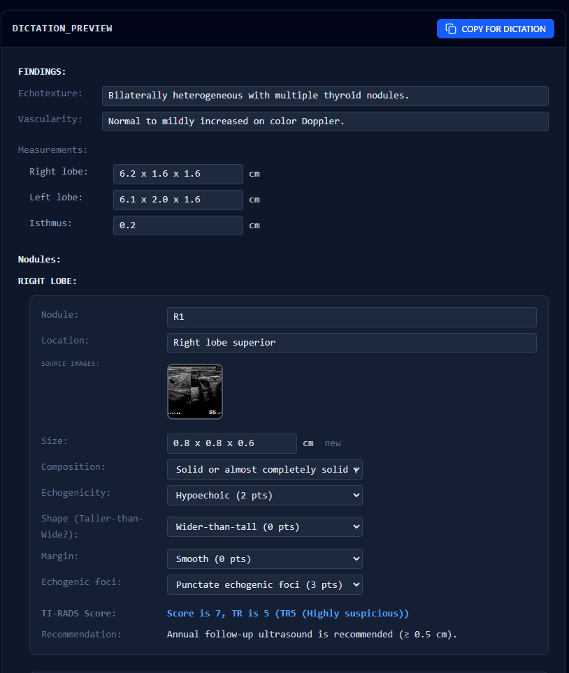
The real interface: structured dictation preview, every field mapped to your EHR schema
Recognized By
Startup Program Members

Infrastructure & Compute Partners
GPU Compute · Medical Imaging · AI Acceleration
Cloud AI · Infrastructure · Compute
HIPAA Cloud · Secure Storage

Dr. Mo Fakhri, MD
Founder & CEO · Practicing Neuroradiologist
“I read these studies every day. I code the system that reads them. There is no translation layer between clinical insight and engineering.”
Clinical Workflow Add-On
Voxel Suite: The Workflow Layer Around the Reads
Ships alongside the AI reads: matched priors and their impressions, reason for exam, clinical history, and CT radiation dose, gathered and pasted into your dictation software automatically. Reduces dictation time by up to 80% (internal estimate), with no AI interpretation involved.
Ready for drafts across your whole worklist?
25+ exam types across X-ray, ultrasound, and CT, drafted before the study is opened and signed by your radiologists. Request early access to Voxel Vision.